Macular degeneration
A blurred or dark patch in the centre of vision, more common after 50.


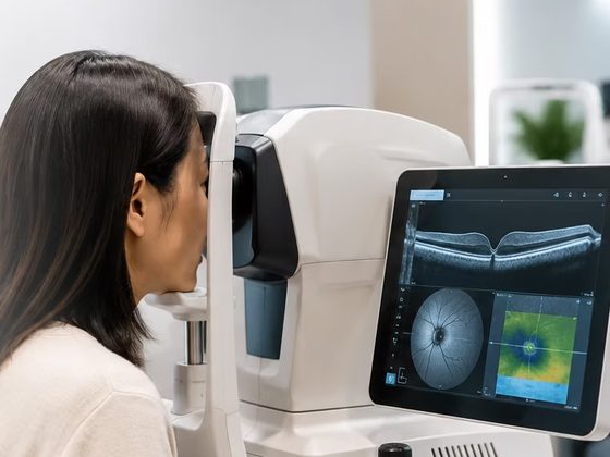
A cross-section of the eye. An OCT scan images the retina and optic nerve at the back of the eye, and the front of the eye too. Nothing touches the eye.
Camden (Orchard)Joo ChiatClementiBedokToa PayohJurong
Found this helpful?

OCT stands for optical coherence tomography. It is a scan that images the eye in cross-section, using light rather than sound or X-rays.
You will also see it written as ocular coherence tomography, and asked for as an OCT eye test, an OCT eye examination or an OCT eye scan. All of those mean this one test. Optical is the correct word, because it is light the scanner works with.
A beam of light is scanned across the retina


Been told you need a scan? Tell our friendly staff what you have been told and they will arrange the right appointment.
What you may have noticed, and the conditions the scan helps the doctor look at.
A blurred or dark patch in the centre of vision, more common after 50.
Straight lines look wavy or bent, or print blurs in the middle.
Swelling at the centre of the retina, which can come with diabetes.
Raised eye pressure, or an optic nerve that needs a closer look.
A dim, blurred or distorted spot in the centre of vision.
Some, such as hydroxychloroquine for lupus or arthritis, are checked for their effect on the retina.
If you have a concern, talk to our friendly eye doctors for peace of mind. They will decide whether a scan is needed.
The centre of the retina, and the part of it you read with.
A macular OCT, or an OCT scan of the retina, images the layers under the middle of your vision.
Wavy straight lines are one of the things the Amsler grid checks before a scan is arranged.
Also written as an OCT RNFL scan, after the layer it measures.
The nerve fibres from across the retina leave the eye together at the optic nerve head. The layer they travel in is the retinal nerve fibre layer, RNFL for short.
What a reading means, and what is done about it, is set out under glaucoma and high eye pressure.
Also written as an anterior segment OCT.
The same scan, aimed at the front of the eye rather than the back.
The scanner shines a beam of low-power light into the eye and measures what comes back from each layer. Light returning from a deeper layer takes fractionally longer to come back, and it is that difference the machine turns into depth.
Drops to widen the pupil are not always needed for an OCT scan. Whether they are used is decided at the visit, and they blur near vision for several hours if they are.

Both look at the retina. One is flat and one is a cross-section, and that is the whole difference.
| What is compared | Retinal photograph | OCT scan |
|---|---|---|
| What it produces | A flat picture, the way a camera sees the retina. | A cross-section, layer by layer. |
| How it is made | One flash of light through the pupil. | A beam of light scanned across, measuring what each layer reflects back. |
| What it is for | Colour, and the pattern across the whole retina. | Depth and thickness, at the spot being scanned. |
Neither replaces the other, so both are often done at the same visit, and either may be called a retinal scan.
Our screening packages carry two kinds of OCT scan, and they scan different ends of the eye.
The screening packages are optometrist-led and every report is reviewed by an ophthalmologist.
| Screening package | OCT included | Price |
|---|---|---|
| General eye screening | Front of the eye | $99 |
| Diabetic retinal screening | Front of the eye | $99 |
| Comprehensive eye screening | Front of the eye and retina | $249 |
Prices shown are for our community clinics at Bedok, Clementi, Toa Payoh and Jurong. All fees shown are nett, and each figure is the price of the whole screening package, not of the scan on its own.
A scan can also be done at a consultation, which is the route for an optic nerve scan and for one repeated over time. No price is published for a scan on its own, so ask our staff when you book.
Optical coherence tomography. A scanner sends a beam of light into the eye and measures what each layer of tissue reflects back, then builds those measurements into a cross-section image of the retina, the optic nerve or the front of the eye. Nothing touches the eye and there is no injection.
No, and yes. You rest your chin and forehead against a frame and look at a target while the scan runs. The light is bright for a moment, but nothing touches the eye, there is no needle and no dye, and an OCT scan uses light rather than X-rays, so it involves no radiation.
The scan itself takes a matter of seconds for each eye. Allow a few minutes at the machine for being positioned and for the images to be checked before you get up. If dilating drops are used, they add 15 to 30 minutes before the scan while they work.
If no dilating drops were used, yes — the scan itself does not change your vision. If your pupils were dilated for it, near vision is blurred and bright light is uncomfortable for several hours afterwards, sometimes longer, so arrange not to drive.
A retinal photograph is a flat picture of the surface of the retina, the way a camera sees it. An OCT scan images the same tissue in cross-section, so it shows the thickness of each layer rather than the pattern across the whole retina. They answer different questions and are often done at the same visit.
A scan of the retinal nerve fibre layer, the layer of nerve fibres that gathers at the optic nerve and leaves the eye there. The scan measures how thick that layer is around the nerve head. It is done as part of a consultation, and the reading is read alongside the eye pressure and the visual field rather than on its own.
No price is published for an OCT scan on its own. At our community clinics in Bedok, Clementi, Toa Payoh and Jurong, a scan of the front of the eye is part of the $99 general and $99 diabetic retinal screenings, and the $249 comprehensive screening adds a scan of the retina. For a scan at a consultation, ask our staff when you book.
Send us a message with what you have been told and a time that suits, and our staff will arrange the appointment.