
Common Eye Tests
What each test is for. From the letter chart to a scan of the retina, what each one looks at, how it is done, and which of them use drops.
- Senior Consultant Eye Surgeons
- 50+ combined years in ophthalmology
- Seven clinics in six locations islandwide
Camden (Orchard)Joo ChiatClementiBedokToa PayohJurong
Found this helpful?

What your eye tests are for
An eye examination is a short series of tests, each looking at a different part of the eye: how you see, the surface, the pressure inside, the retina and your side vision.
These are the common ones. We can also arrange more advanced tests if needed. If you have a concern, talk to our eye doctors.
- Vision and prescription What you can see, and what glasses would change. 3 tests
- The front of the eye The lids, the cornea, the iris and the lens. 4 tests
- Eye pressure The pressure inside, and the drainage angle. 3 tests
- The back of the eye The retina, the macula and the optic nerve. 4 tests
- Side vision The vision around what you look at. 1 test
- In a screening The tests every screening package includes. 4 packages


Not sure what you need? Tell our friendly staff what you have noticed and they will arrange the right appointment.
Vision and prescription
The tests that measure what you can see, and what glasses would change about it.
-
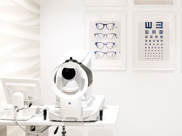
Visual acuity
How clearly you see, read off a Snellen letter chart one eye at a time. The result is written as a pair of numbers, 6/6 being the standard line.
In every screening package
-
Automated refraction
A machine estimate of your spectacle prescription. It is a starting point rather than the final figure, which is settled by checking lenses against your own vision.
In every screening package
-
Colour vision test
Plates of coloured dots with a number hidden inside them, which is how a colour vision deficiency is checked.
In two screening packages
Visual acuity is not the same thing as your prescription. It says how much you can see today, with or without the glasses you already own; the refraction says what lenses would be needed to change it.
The front of the eye and the tear film
The lids, the cornea, the iris and the lens, at magnification.
-
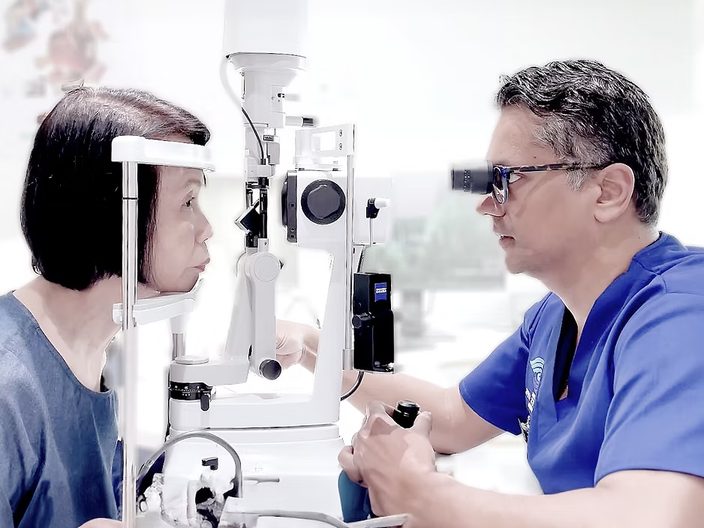
Slit lamp examination
A microscope with a bright, narrow beam of light. You rest your chin and forehead against a frame and the beam is swept across the eye, which gives a magnified view of the lids, the cornea, the iris and the lens behind them.
In every screening package
-
Fluorescein stain
A drop of orange dye, looked at under a blue light. The dye settles where the surface of the cornea is broken or dry, so those areas can be seen.
At a consultation
-
Tear break-up time
Measured with the same dye: you blink once and the seconds are counted until the film of tears over the cornea starts to break up.
At a consultation
-
Anterior segment photographs
A photograph of what the slit lamp shows, so the same eye can be compared with itself at a later visit.
At a consultation
The slit lamp is the test most people have without knowing its name. It is in all four of our screening packages, and it is how the lens itself is looked at — which is where a cataract is found.
Eye pressure and the drainage angle
Three tests that are read together, because each one changes how the others are understood.
-
Tonometry
A measurement of the pressure inside the eye, in millimetres of mercury. Some methods puff air at the eye, others touch it lightly after an anaesthetic drop.
In every screening package
-
Central corneal thickness, or pachymetry
A measurement of the cornea. A thick cornea can make a pressure reading look higher than it is and a thin one lower, so the two are read as a pair.
At a consultation
-
Gonioscopy
A small mirrored lens rested against a numbed eye, which lets the drainage angle be seen — the corner where fluid leaves the eye, and one that cannot be seen looking straight on.
At a consultation
One pressure reading on its own is a number, not a diagnosis: it varies through the day and between people. What counts as high, and what is done about it, is set out under high eye pressure and glaucoma.
The back of the eye
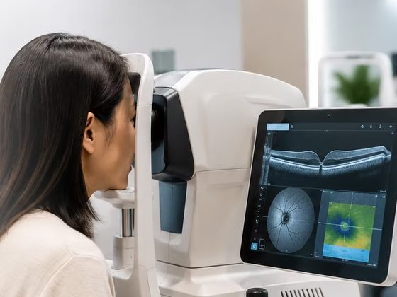
The retina, the macula at its centre, and the optic nerve leaving it.
-
OCT scan
A scan that images the retina in cross-section, layer by layer, using light rather than sound or X-rays. It shows depth, which a photograph cannot.
In the Comprehensive screening
-
Fundus photograph
A photograph of the retina, taken through the pupil. It records the optic nerve, the macula and the blood vessels as a flat image, the way a camera sees them.
In three screening packages
-
Amsler grid
A square grid with a dot in the middle. You look at the dot and say whether the lines around it look straight or complete, which is a check of the central vision the macula gives you.
In two screening packages
-
Dilated examination
Drops widen the pupil so more of the retina can be examined through it. They take 15 to 30 minutes to work and leave near vision blurred and light uncomfortable for several hours afterwards, sometimes longer, so it is worth arranging not to drive.
In the Diabetic retinal screening, where necessary
A photograph and a scan answer different questions about the same tissue, and are often done at the same visit.
Side vision
The vision around what you are looking at, rather than the vision you read with.
-
Visual field test
You look steadily at a central point inside a bowl-shaped screen and press a button each time a small light appears somewhere in the field around it. One eye is covered at a time and each eye takes a few minutes. The result is a map of where lights were seen and where they were missed.
It is tiring rather than uncomfortable, and it is normal to miss some lights: the map is read as a pattern, not as a score.
In the Comprehensive screening
Which tests come with an eye screening
All four of our screening packages include the same four tests, and every report is reviewed by an ophthalmologist.
- Visual acuity How clearly you see on a Snellen letter chart.
- Automated refraction A machine estimate of your spectacle prescription.
- Eye pressure check Tonometry — measurement of the pressure inside the eye.
- Slit lamp examination A magnified look at the front of the eye.
The packages differ in what they add on top of those — a retinal photograph, a colour vision test, a scan of the front of the eye, and in the fullest package, side vision testing and a scan of the retina. Which tests sit in which package is set out in full on the screening pages, package by package.
Prices shown are for our community clinics at Bedok, Clementi, Toa Payoh and Jurong. A test can also be done as part of a consultation, where which ones you have is decided by the doctor seeing you.
Common questions
Why do I need so many eye tests?
Each one looks at a different part of the eye, and no single test covers all of it. A letter chart measures how clearly you see but says nothing about the pressure inside the eye; a pressure reading says nothing about the retina. Which tests you have depends on what you have come in about, your age and your medical history.
Which eye tests use drops?
Three kinds. Anaesthetic drops numb the surface for tests that touch the eye, such as gonioscopy. An orange fluorescein dye is used to look at the surface of the cornea and the tear film. Dilating drops widen the pupil for a fuller view of the retina, and they blur near vision for several hours afterwards, sometimes longer. Of our four screening packages, only the diabetic retinal screening dilates, and only where it is necessary.
What is gonioscopy?
A look at the drainage angle, the corner where the iris meets the cornea and where fluid leaves the eye. The eye is numbed with a drop and a small mirrored lens is rested against it, which lets the doctor see a corner that cannot be seen straight on. It takes a minute or two and is not painful.
What is a corneal thickness test?
A measurement of how thick the cornea is, also called pachymetry. It matters because a thick or a thin cornea shifts an eye pressure reading, so the two numbers are read together rather than one on its own.
Book an eye examination
Send us a message with what you have noticed and a time that suits, and our staff will arrange the appointment.